Electron Microscopy, as we know it today, had its origins in the 1930s and was immediately recognized as a powerful research tool for the visualization of cellular structures at a much higher magnification and degree of resolution than methods currently available to the researcher.
It quickly became apparent that in addition to encompassing the new, enhanced and specialized specimen preparation techniques required for this higher resolution imaging process, the production of ultra-thin sections of biological material suitable for examination in the hostile environment of the electron microscope was important.
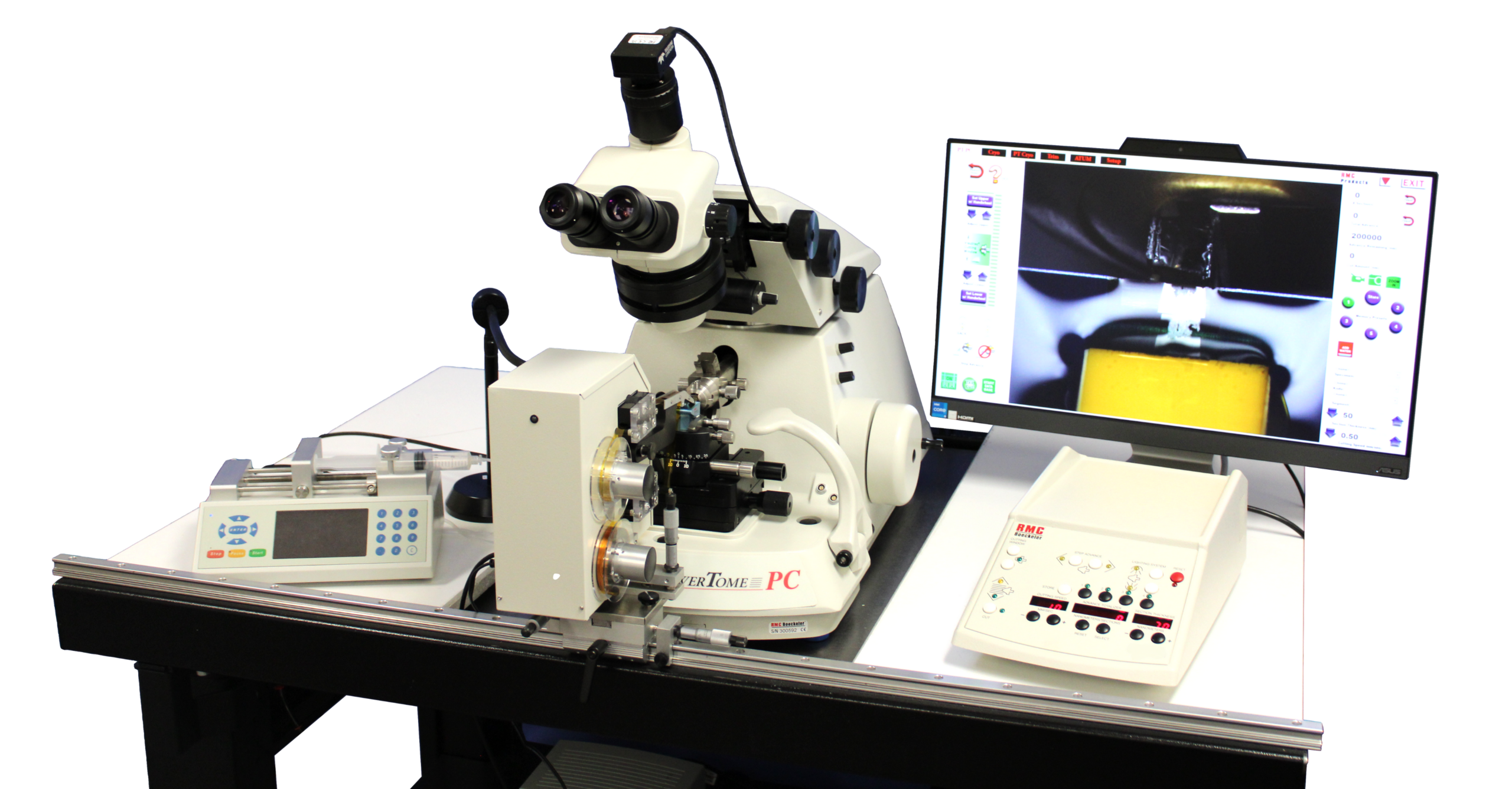
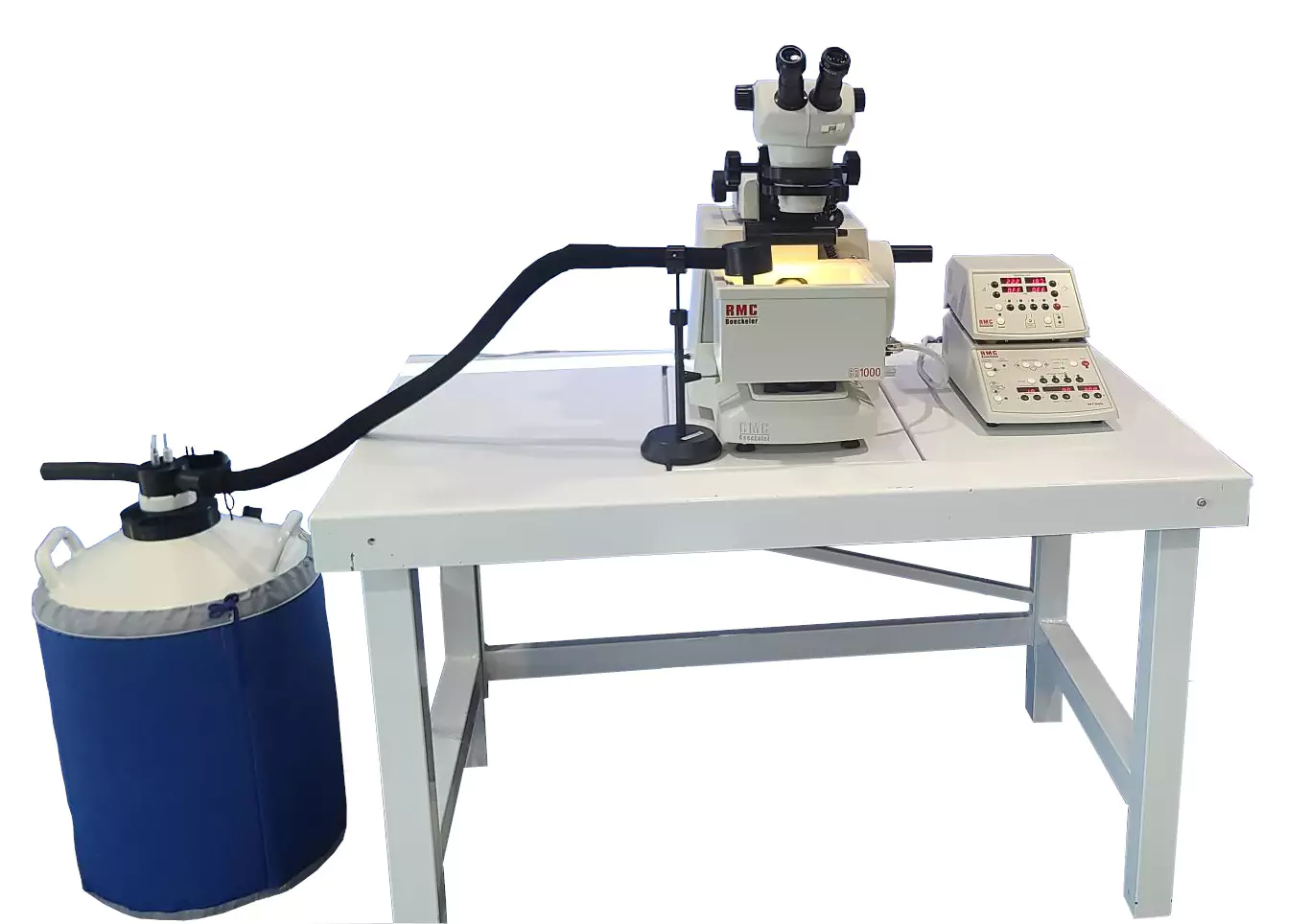
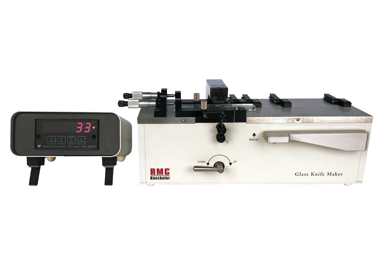
The technique of microtomy was well understood whereby thin sections (typically 3.0 – 10.00um) of a prepared sample were cut using extremely sharp metal knives on a specialized instrument that gave consistent and reproducible results; the microtome. For electron microscopy, a new generation of microtome, the ‘ultra-microtome’ needed to be developed. This instrument needed to be capable of making super thin sections (typically 60 – 100nm) of a sample embedded in a much harder and more resilient medium than previously employed. This new instrument must also display reliability, accuracy, reproducibility, and, of course, ease of use. Many groups or individual engineers endeavored to produce such an instrument.
In 1953, scientist Keith Porter and mechanical engineer Joseph Blum, from the Rockefeller Institute, designed and developed the first commercially successful ultramicrotome in the world that combined the above characteristics. This instrument, the Porter-Blum MT1 (Microtome One) was manufactured and marketed by Sorvall Inc. The direct descendent of this groundbreaking instrument is today manufactured and marketed by Boeckeler Instruments Pte Ltd, under the RMC Boeckeler brand, thus maintaining a proud tradition.